baby chest x ray technique
You will be told to hold your breath when the x-ray is taken. Radiographs of the chest and the abdomen are the most commonly requested diagnostic X-ray examinations undertaken in neonatal intensive care units.

Chest Radiograph Pediatric Radiology Reference Article Radiopaedia Org
Up to 5 cash back Chest and Abdomen Stephanie French Senior Radiographer Leeds General Infirmary.

. Chest Routine chest. A chest x-ray is an x-ray of the chest lungs heart large arteries ribs and diaphragm. From the back of the chest if the child is old enough to stand up for the X-ray from the side.
1A Chest radiographs of two different patients. 200-245 millirad for an x-ray of the abdomen. 51-370 millirad for x-rays of the hip and femur thighbone It is very rare for a single diagnostic x-ray to exceed even 5 rad.
X-rays are used throughout the body. Two images are usually taken. Initial chest radiograph obtained immediately after surgery A shows normal contour of superior mediastinumFollow-up chest radiograph obtained.
PA or AP and left lateral supine in infants upright in children or when requested. Chest X-Ray - Lung disease. It is often the first type of imaging used to identify sources of pain evaluate traumatic injuries and locate a foreign body.
You would have to x-ray your arm or leg more than 5000 times in order to reach 5 rad of exposure to your unborn baby. Lateral cervical spines are taken at 150 cm. Chest X-rays can be done while a child is standing sitting or lying down.
Chest X-ray - Heart Failure. Your child may be required to hold his or her breath or remain still. Chest x-ray is the most commonly used imaging exam for evaluating the chest.
It can help diagnose and assess. Frequently for a single child both radiographs are requested simultaneously. Most neonatal chest X-rays are AP films unless the baby is made to lie prone Lucency of soft tissue shadow - darker the soft tissue more is the exposure Ease of visibility of retrocardiac vertebrae if the retrocardiac vertebrae are easily seen the film is over exposed Relative lucency of lung fields.
Birth Weight Gestational Age and Vaginal or Cesarean. For younger children the technician will take pictures from the front of the chest and the side. 32 cases of suspected COVID-19.
Baby chest x ray technique. AP supine and lateral cross-table o Newborn Initial Chest. Sometimes other picture views also are taken.
The mean effective dose for the separate exposure technique was estimated to be 373 microSv compared with 355. The FDA recommends that medical x-ray imaging exams which include computed tomography CT fluoroscopy and conventional X-rays use the. Once the radiographer a person specially trained in taking X-ray images has positioned the part of your childs body to be examined and lined up the X-ray machine the X-ray examination takes less than a second to perform.
You stand in front of the x-ray machine. If exam is the initial chest image please include the following in tech comments. The chest radiograph is the most common radiographic procedure performed in the imaging department and is the initial imaging modality in a patient presenting with thoracic symptoms.
Erect chest X-rays are taken at 180 cm. X-ray examinations are usually quick and simple. How the Test is Performed.
Chest Abdo UAC - The tip is overlying. It would take more than 50 chest x-rays to reach 5 rad. Usually the X-ray technician will take pictures of the chest.
Baby must be prone 20mins prior to exposure. X-ray exams are used to help diagnose a wide variety of injuries and illnesses in children. In this study the exposure technique of 65 kVp and 16 mAs was chosen as a reference image due to this technique being near the suitable exposure uses in pediatric chest supine AP in the DR and.
Chest X-Ray - Basic Interpretation. All distal extremity exposures are taken at 110115 cm SID. Full legfull spine imaging is performed at 180 cm using CR.
- Use good technique - Promote optimal image quality - Ensure that the infant is not exposed to any minimal environmental stress as a result of increased handling and. A 9-month-old girl with history of atrioventricular septal defect who underwent repair and heart block requiring permanent pacemaker now receiving warfarin. X-ray plate - avoid direct contact with baby use X-ray tray provided with the open care system cover with sterile plastic sheetenvelope Avoid direct contact with the cold X-ray plate Provide extra heat source as the open care heater is moved away from the baby or tilt its direction towards baby if possible.
Make use of digital radiography dr and needle phosphor computerised.

Chest X Ray Of A 6 Month Old Child With An Icd The Active Can Is Download Scientific Diagram

Pin By Heather Royal On School Daze Diagnostic Imaging Med Student Medical Field
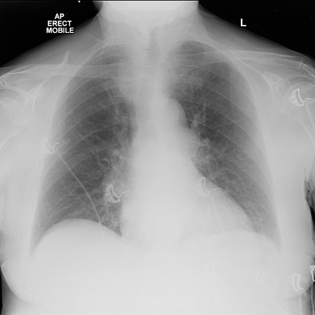
Chest Ap Erect View Radiology Reference Article Radiopaedia Org

Neonate Chest Supine View Radiology Reference Article Radiopaedia Org
2
2
Radiodensity On Serial Chest X Rays For The Diagnosis Of Foreign Body Aspiration In Children

Good Lateral Elbow Diagnostic Imaging Radiology Imaging Radiology Schools

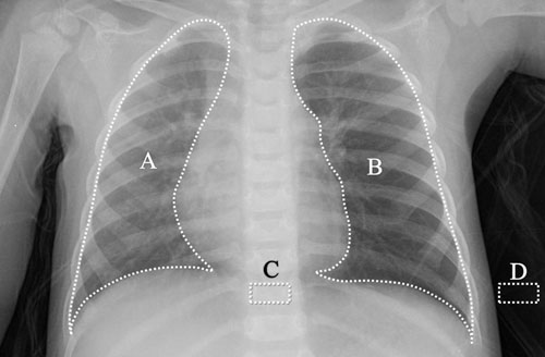
Pediatric Chest Supine View Radiology Reference Article Radiopaedia Org

Chest X Ray Child Youtube

Special Investigations In Cardiology Radiology And Electrocardiography Ecg Radiology Radiology Student Medical Assistant Student

Magnets May Pull Kids With Sunken Chests Out Of Operating Room Pectus Excavatum Chest Marfan Syndrome
Approach To Pediatric Chest X Rays Youtube

Normal Chest X Ray Of 7 Year Old Girl Stock Photo Alamy

Pedia Poser For Xray Imaging

A Pediatric Chest X Ray With The Pb Shield The Circles Are The Download Scientific Diagram

Fmes Net Chest X Ray Analysis

Ce4rt Guide For X Ray Techs To Immobilize Pediatrict Patients
2